Smartphone imaging tool shows promise for early oral cancer detection
New smartphone imaging system shows 60% sensitivity for oral cancer lesions; could assist dentists in low-resource settings.
New device captures oral lesions with dual imaging
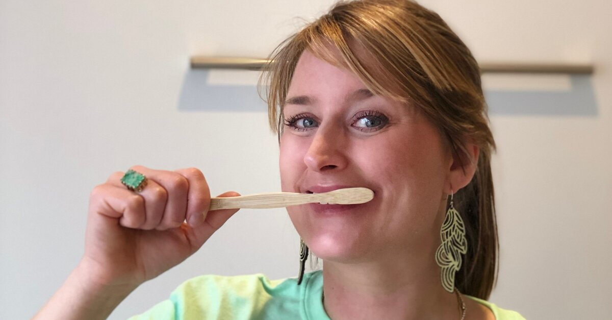
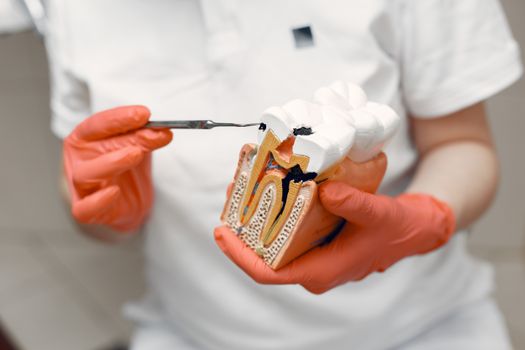
A smartphone-based imaging system called mDOC has been evaluated for its ability to help dentists identify suspicious oral lesions early. The system captures both white light and autofluorescence images of the oral cavity, revealing tissue changes not visible under standard lighting. It also collects patient risk factors such as age, smoking history, and lesion location, then uses this information to recommend whether specialist evaluation is needed. The imaging process takes approximately 3.5 minutes, making it feasible to integrate into routine dental visits.
Detection performance and clinical limitations
In a study of 50 patients at UTHealth Houston School of Dentistry and Harris Health Dental Center, the mDOC system was compared with assessments from dental hygienists, general dentists, and expert clinicians. The system detected more potentially dangerous lesions than the dentists and hygienists examined without imaging support. However, it correctly identified only 60% of lesions that experts recommended for referral, showing moderate sensitivity. The system produced several false positives, indicating room for improvement. Notably, unaided clinicians in the study identified none of the cases requiring referral, suggesting that adjunctive imaging tools may enhance detection in routine practice. The researchers emphasize that the system should complement, not replace, clinical judgment.
Potential for underserved communities
According to lead author Dr Ruchika Mitbander, early oral cancer detection is essential because survival rates decline as the disease progresses. In community dental settings, particularly in underserved or low-resource regions, providers often lack reliable tools for identifying lesions requiring specialist referral. The mDOC system offers a portable, dual-mode imaging device that integrates multiple data inputs into a single decision support tool. Beyond screening, it may support monitoring of existing lesions and could be combined with additional diagnostic methods such as cytology in settings with limited specialist access. The study was published online on 6 October 2025 in Biophotonics Discovery.
Frequently asked questions
What is the mDOC system and how does it work?
mDOC is a smartphone-based imaging system that captures paired white light and autofluorescence images of the oral cavity to visualize tissue changes not apparent under conventional lighting. It also collects patient risk factors such as age, smoking history, and lesion location, then analyzes this data to recommend whether specialist evaluation is needed. The entire imaging process takes approximately 3.5 minutes.
What was the sensitivity of the mDOC system in the study?
The mDOC system correctly identified 60% of lesions that expert clinicians recommended for referral. It detected more potentially dangerous lesions than unaided dentists and hygienists, but also produced a number of false positives, indicating the need for further optimization before clinical deployment.
Can the mDOC system replace clinical judgment by dentists?
No. The researchers emphasize that the mDOC system should complement, not replace, clinical judgment. It is intended as an adjunctive decision support tool to help dentists identify lesions that warrant specialist referral, particularly in community dental settings where training in lesion recognition may be limited.
Why is this system useful in underserved dental settings?
In underserved or low-resource regions, dental providers often lack reliable tools for identifying oral lesions requiring specialist referral. The portable mDOC system provides an adjunctive screening tool at the point of care, potentially bridging the gap between routine screening and specialist access, and helping improve outcomes in communities with limited specialist availability.